Originally published on Ultrasound G.E.L. on 4/10/17 – Visit HERE to listen to accompanying PODCAST! Reposted with permission.
Follow Dr. Michael Prats, MD (@PratsEM), Dr. Creagh Bougler, MD (@CreaghB), and Dr. Jacob Avila, MD (@UltrasoundMD) from Ultrasound G.E.L. team!
Accuracy of Point-of-care Lung Ultrasonography for Diagnosis of Acute Chest Syndrome in Pediatric Patients with Sickle Cell Disease and Fever
Academic Emergency Medicine August 2016 – Pubmed Link
Take Home Points
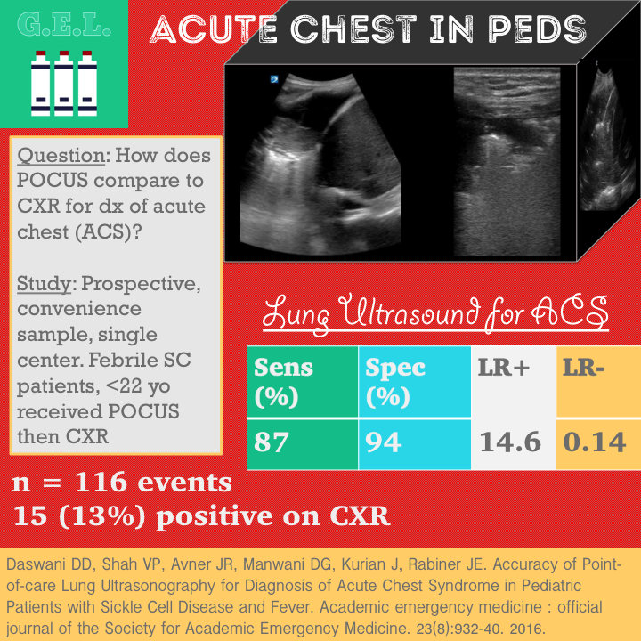
1. POCUS is 94% specific and 87% sensitive for acute chest syndrome in pediatric patients when compared to chest xray.
2. In certain circumstances, the combination of a high pretest probability and a positive lung ultrasound may be sufficient in ruling in acute chest syndrome.
3. In this study, operator experience significantly affected the accuracy of the ultrasound.
Background
Acute chest syndrome (ACS) is a major cause of mortality in sickle cell anemia disease (SCD). About one third of patients with SCD have it in their life time. 17% of sickle cell patients with fever will have ACS. Unfortunately, the physical exam lets us down again – half of these patients have no physical findings. The diagnosis is usually made by variations of this formula: history of sickle cell disease + symptoms consistent with acute chest syndrome + new infiltrate on chest xray = ACS. Obviously, this is not fool-proof but it is more or less how the initial diagnosis takes place. As a consequence, patients with SCD will have an average of 27 radiographic tests by age 18. Maybe there is another option without radiation? We already know that ultrasound is pretty good at diagnosing pneumonia in pediatrics (see our prior post and podcast here) and this is similarly a consolidative process so it seems like a reasonable idea.
Questions
How accurate is point of care lung ultrasound in diagnosing ACS?
Secondary objective is to evaluate the interobserver agreement between pediatric emergency medicine (PEM) physicians and an experienced PEM sonologist
Population
Urban pediatric ED
August 2013 to February 2015
Inclusion
- up to 21 years old
- diagnosis of SCD
- fever (>38 C) within 24 hrs
- requiring chest xray
Exclusion
- CXR within past 5 days for fever (consider part of ongoing illness)
- diagnosis of ACS within previous 6 weeks (may have residual radiographic findings).
- not excluded if already participated in study
Who did the scans?
PEM physicians
1 hour didactic and hands-on training on US of lung
One on one hands on training with live adult and peds models
Design
Prospective, observational study
Convenience sample – if presenting when trained PEM physician available
Intervention
All patients got institutional protocol for suspected ACS:
- labs
- cxr
- IV antibiotics
- phone consultation with hematologist
Prior to imaging, history and exam findings were recorded. A PEM physician recorded likelihood of ACS into one of 6 categories of percentage of likelihood.
Before cxr, point of care ultrasound was performed by a second PEM physician who was blinded to clinical info and physical exam (they did know it was a sickle cell patient with fever, and they could see the patient for any observable findings).
Comparison was CXR as read by attending radiologist, blinded to US
Equivocal reads were reviewed by a second radiologist
Follow up was performed by review of records and/or telephone follow up (asking about additional visits, imaging, or admissions since first ED visit)
The Scan
Linear probe

6-zone lung (bilateral anterior, lateral and posterior)
Clips, images, long and short axis
Positive LUS = consolidation with bronchograms. Consolidation = hepatization, loss of a-lines, disruption of hyperechoic pleural line
Scanning for acute chest is the same as scanning for pneumonia – see the findings here!
Results
91 patients, 116 events
- Median age 5.7 years, Range 4 months to 20 years
- Follow up obtained in 99.1% of patients (all still included in analysis)
- 3 patients who had no ACS on initial visit were diagnosed within 7 days (6 and 7 days later). This speaks to the accuracy of chest xray in making the diagnosis.
CXR positive 13% (15 cases) Mostly consolidations, one with small bilateral pleural effusions that was counted)
Lung ultrasound (LUS) positive 16% (air bronchograms with consolidation)
Primary Outcome
Lung Ultrasound for Acute Chest Syndrome
Sensitivity 87% (62-96)
Specificity 94% (88-97)
+LR 14.6 (6.5-32.5)
-LR 0.14 (0.04 – 0.52)
(similar if excluded repeat visits – sens 85%, spec 95%)
Other Findings
Agreement was kappa of 0.77 between PEM and expert PEM sonologist
In all cases where cxr and LUS was positive – it was same location
Took a median of 9 minutes to perform (for ballpark comparison – a separate study showed average cxr time of 27 minutes)
Physical exam and pre-test probability were not that good for predicting ACS
- Almost half (46%) of the patients with ACS were originally rated as having low likelihood of having the disease
15 PEM physicians, median of 4 scans each, BUT one provider did 36 exams (31%) and had 100% sens and spec → if you exclude those (all other PEM sonologists had <25 LUS scans) sens 82%, spec 81%
2 false negatives (both right upper lobe)
- one was adjacent to thymus (identified on expert review)
- second missed had limited clips to review
6 false positives
- 4 were operator error (spleen + air in stomach diagnosed as consolidation).
- 1 was also likely similar area (diagnosed in left lung base) but no clips saved.
- 1 was confirmed consolidation (missed by xray), treated as ACS on inpatient unit because of symptoms and respiratory distress.
Limitations
Single center, convenience sample
Small population especially with regard to the number of positive exams (only 15 cases of acute chest)
CXR used as gold standard which is a clinically appropriate comparison but likely does not reflect true accuracy of disease diagnosis
Broad range of operator accuracies (100% to 80% based on experience). This is likely more dramatic because of the small sample size, but is still concerning because whether it is a useful test or not depends on how good the sonologist is with ultrasound (so get better with ultrasound).
Blinding – good but not perfect (could see desaturation, distress, etc) – but only 1 of these had ACS on LUS and confirmed on CXR (so maybe didn’t bias too much)
Take Home Points
1. POCUS is 94% specific and 87% sensitive for acute chest syndrome in pediatric patients when compared to chest xray.
2. In certain circumstances, the combination of a high pretest probability and a positive lung ultrasound may be sufficient in ruling in acute chest syndrome.
3. In this study, operator experience significantly affected the accuracy of the study.
Our score

The post <div class="qa-status-icon qa-unanswered-icon"></div>Ultrasound G.E.L. – Acute Chest Syndrome in Pediatric Patients appeared first on emDOCs.net - Emergency Medicine Education.
